 | ||||

Tasmanian Devil
Nearly all types of bacteria have a cell wall containing chemical components that are unique to bacterial cells. The rigid structure of the bacterial cell wall is due to securely linked peptidoglycan molecules that surround the plasma membrane, giving these prokaryotic cells shape and protection.
Gram- Cell Wall
Peptidoglycan: From the peptidoglycan inwards nearly all bacterial cells are very similar. Going further out, the bacterial world divides into two
major groups:
Article Summary: Most bacteria have one of two types of cell walls. Here are the features of the Gram- bacterial cell wall that distinguish it from Gram+.

Gram-negative, lactose fermenting
SPO VIRTUAL CLASSROOMS
Gram positive (Gram+) and Gram negative (Gram-). The cell walls of Gram - bacteria are more chemically complex, thinner and less compact, with peptidoglycan comprising only 5 – 20% of the cell wall.
LPS Membrane: In gram-negative bacteria, peptidoglycan is not the outermost layer of the cell wall. Gram- cells have an additional, external membrane, similar to the plasma membrane, but less permeable and composed of lipopolysaccharides (LPS); a harmful substance classified as an endotoxin.

Gram-negative Escherichia coli @ 1000xTM
The Gram Stain
Once scientists understood that infectious disease was caused by microorganisms (Germ Theory), it was imperative to find a way to view bacteria and other microbes; because in addition to being minute, most bacteria colorless.
This differential staining not only colors the bacteria, but the specific stain reaction distinguishes between two meaningful categories of bacteria based on the differences in their cell wall structure.
After this staining procedure, Gram + cells appear purple; their thick layers of peptidoglycan having retained the primary stain, crystal violet. Because Gram negative cells have a very thin layer of peptidoglycan layer, these cells do not retain the purple primary stain. At the end of the Gram staining procedure, Gram-negative cells retain the secondary stain, safanin, and appear pink.
Gram Negative Bacteria as Pathogens
Many Gram-negative bacteria are pathogens; bacteria that can cause disease. This pathogenicity is typically associated with lipopolysaccharide (LPS) endotoxins in Gram-negative cell walls, and other Gram-negative virulence factors such as the fimbriae, which help bacteria adhere to cells they can infect.
Sources & Resources
- Bauman, R. (2014) Microbiology with Diseases by Taxonomy 4th ed.
- Park Talaro, K. (2008) Foundations in Microbiology.
- Gram Staining & Isolation Streak Plate Laboratory Main Page from the Virtual Microbiology Classroom
- Differential Staining of Bacteria Laboratory Main Page.
- Gram Stain Bite Sized Tutorial: This is an extremely useful tutorial that shows, step-by-step, the Gram-staining procedure and the appearance of Gram+ and Gram- bacterial cells.
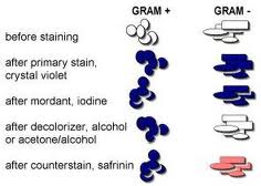
In the 1800’s, Christian Gram, a Danish bacteriologist, developed a technique for staining bacteria that is still widely used today. The Gram stain protocol involves the application of a series of dyes that leaves some bacteria purple (Gram +) and others pink (Gram -).

SCIENCE PHOTOS


Continued ...
Page last updated: 11/2015
You have free access to a large collection of materials used in a college-level introductory microbiology course. The Virtual Microbiology Classroom provides a wide range of free educational resources including PowerPoint Lectures, Study Guides, Review Questions and Practice Test Questions.


H. C. Gram
