 | ||||
How to Do a Gram Stain
Test for Gram+ & Gram- Bacteria Identification
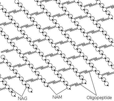
Peptidoglycan is a huge polymer of interlocking chains of NAG and NAM polysaccharide monomers connected by interpeptide bridges.
Bacterial Cell Wall: Peptidoglycan Structure
This rigid structure of peptidoglycan gives the bacterial cell shape, surrounds the plasma membrane and provides prokaryotes with protection from their environment.
Article Summary: Gram staining involves the application of a series of dyes that leaves some bacteria purple (Gram +) and others pink (Gram -). Here's how the Gram stain works.
Gram Stain for Identifying Gram +/- Bacteria
Page last updated: 11/2015
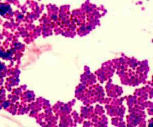
Gram-positive
Staphylococcus epidermidis @ 1000xTM
SPO VIRTUAL CLASSROOMS
 | ||||||
How to Prepare a Bacterial Smear
for Gram Staining
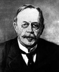
In the 1800’s, Hans Christian Gram, a Danish bacteriologist, developed a technique for staining bacteria that is still widely used today.
The Gram stain protocol involves the application of a series of dyes that results in some bacteria staining purple and others pink.
Bacteria that stain purple are considered Gram-positive, and those that stain pink, Gram-negative. The specific stain reaction of a bacterium results from the structure of the bacterial cell wall.
You have free access to a large collection of materials used in a college-level introductory microbiology course. The Virtual Microbiology Classroom provides a wide range of free educational resources including PowerPoint Lectures, Study Guides, Review Questions and Practice Test Questions.
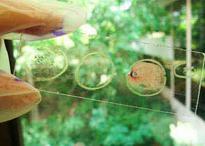
A smear is a sample of bacteria suspended in a small amount of water on a slide. That sample is then dried using heat. The heat kills the bacteria and attaches the sample to the slide so that it does not easily wash away.
From the peptidoglycan
inwards all bacterial cells are very similar. Going further out, the bacterial world divides into two major classes: Gram positive (Gram+) and Gram negative (Gram-).
H. C. Gram
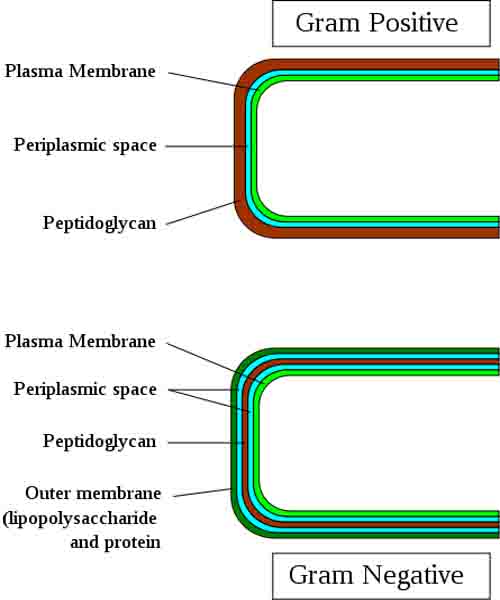
Gram-positive Cells: In Gram-positive bacterial cells, peptidoglycan makes up as much as 90% of the thick, compact cell wall, which is the outermost cell wall structure of Gram+ cells.
Gram-negative Cells: The cell walls of Gram-negative bacteria are more chemically complex, thinner and less compact. Peptidoglycan makes up only 5 – 20% of the cell wall, and is not the outermost layer, but lies between the plasma membrane and an outer membrane. This outer membrane is similar to the plasma membrane, but is less permeable and composed of lipopolysaccharides (LPS), a harmful substance classified as an endotoxin.
Gram Staining Procedure
Because most bacteria have one of these two types of cell walls, we can use this difference as a feature that can be identified using the Gram stain. The Gram stain is a differential stain that uses two dyes to differentiate between the two basic bacterial cell wall types.
First a bacterial smear must be heat fixed to a microscope slide.
