 | ||||
What Is and Electron Microscope?
Basics of Scanning and Transmission EMs
LAB NOTES from Science Prof Online
Science students use light microscopes to view many types of tiny specimens, usually biological cells and small organisms. Light microscopes employ a beam of light to visualize objects. But because light is used, specimens smaller than a wavelength of light cannot be clearly viewed.
Resolution is the amount of detail you can see in an image. You can enlarge an image indefinitely using more powerful lenses, but the image will eventually become blurry. Therefore,
Article Summary: Transmission and scanning electron microscopes use a beam of electrons to magnify and visualize microscopic objects. Here's a comparison of SEMs and TEMS.
Scanning & Transmission Electron Microscopes
 | ||||
The Virtual Microbiology Classroom provides a wide range of FREE educational resources including PowerPoint Lectures, Study Guides, Review Questions and Practice Test Questions.
Page last updated: 10/2014
SCIENCE PHOTOS
SPO VIDEO Tutorial on Compound Light Microscope Parts & Operation
increasing the magnification will not improve the resolution, a.k.a. resolving power.
Light microscopes can generally magnify objects a maximum of about 1000xTM. Microscopic objects and details smaller or closer together than two tenths of a micrometer (0.2 µm)—such as the ultrastructure of eukaryotic cells, detailed structures of bacteria as well as viruses—can only be viewed with the use of an electron microscope.
 | ||||||
SPO VIRTUAL CLASSROOMS
 | ||||
Types of Electron Microscopes
There are two general kinds of electron microscopes—transmission electron microscopes (TEMs) and scanning electron microscopes (SEMs).
Transmission electron microscopes
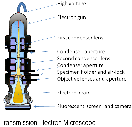
have a tall column topped with an electron gun that fires a beam of electrons downward through a thinly sliced specimen, producing a 2-D image onto a phosphor-coated, fluorescent screen at the bottom of the column. Dense areas of the specimen block electrons, creating darker areas in the image, and less dense areas of the specimen, where more electrons pass through, generate bright areas on the screen. The image that results looks much like that produced by a light microscope, but with much greater magnification. TEMs work well for imaging the internal structures of cells.
More Microscope Resources
- "How big is a...?" animation from Cells Alive.
- Resolving Power Line illustration of what can be seen with different microscopes from Nobelproze.org.
- Microscopic Measurement: How Big Is that Object in the Microscope? from VirtualUrchin.
- Scanning Electron Microscope Basics video from the Virtual Microscope Training Page, produced by the Imaging Technology Group (ITG), Beckman Institute for Advanced Science and Technology, University of Illinois at Urbana-Champaign.
- Preparing a Sample for the Scanning Electron Microscope video produced by ITG.
- Transmission Electron Microscope Animation from Atomic World.
- Transmission Electron Microscope Animated Tutorial from DoITPoMS, University of Cambridge.
- How to Use a Compound Light Microscope: Biology Lab Video Tutorial, SPO YouTube Video.
- How Light Microscopes Magnify Objects and Are Limited by Resolution, Class Notes Article from SPO.
- Microscopic Fresh Water Pond Life Main Page from Kid Science section of Science Prof Online.
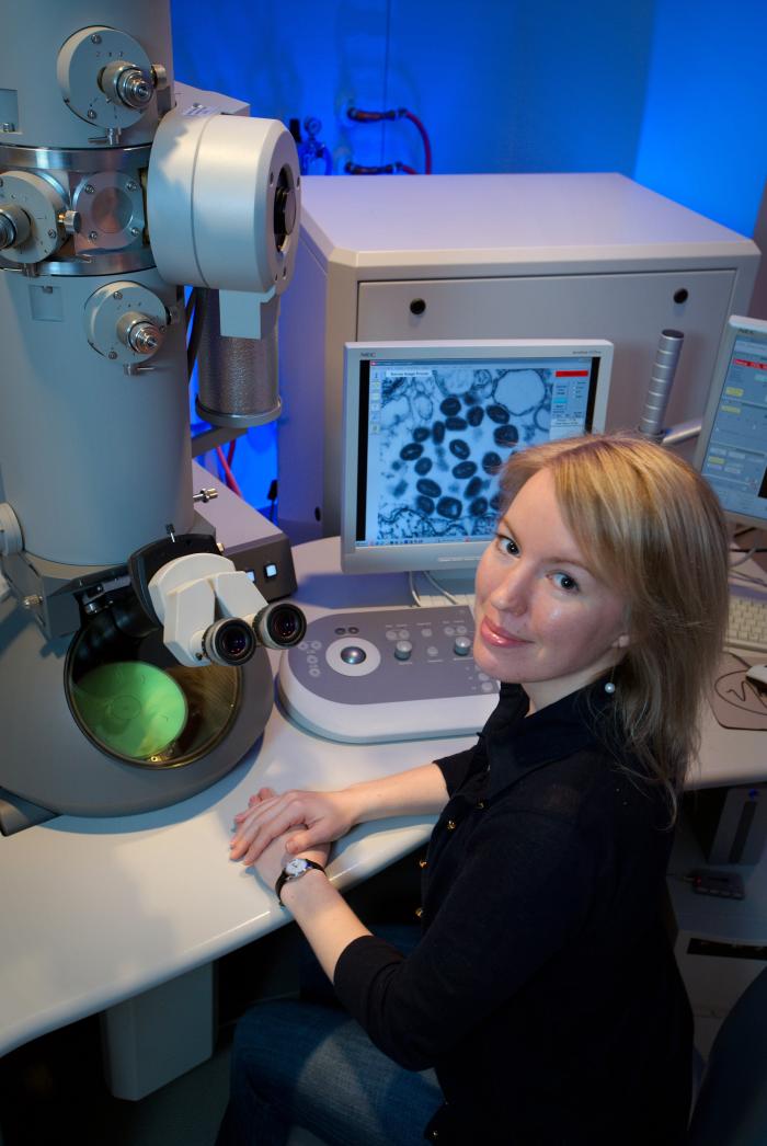
Centers for Disease Control and Prevention (CDC) intern using one of the agency’s transmission electron microscopes (TEM). The microscope’s screen is showing a thin section of the variola virus.
Image of an ant taken with a scanning electron microscope.
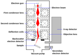
Although light microscopes are relatively small and portable, electron microscopes are usually not. EMs are large instruments, which include an electron gun, the device that shoots a beam of electrons through a vacuum tube into a chamber where the specimen is located.
What Is an Electron Microscope?
Instead of using light, electron microscopes transmit a beam of electrons through, or onto the surface of, a specimen. An electron beam has a much shorter wavelength than does light, and can reveal structures as small as 2 nanometers (nm). For reference, there are 1000 nanometers in 1 micrometer, and 1000 micrometers in a millimeter. So electron microscopes have much more power to magnify. Ultra-high resolution EM can magnify objects 2,000,000x their actual size. However, EMs can only be used to view dead cells, as the method for preparing specimens kills them.
Bullet-shaped rabies virions. Photo taken with transmission electron microscope.
Insect coated in gold, for viewing with a scanning electron microscope.
Scanning electron microscopes
are used to view surface characteristics of a specimen, resulting in a image that reveals three dimensional detail. Rather than penetrating the specimen, the electron beam of an SEM scans and bounces off the surface of the specimen, essentially revealing the specimen’s topography. The resolution of a SEM is about 10 nanometers (nm).
In preparation, the specimen must be thinly coated with metal such as platinum or gold. The electrons from the beam (called primary electrons) knock electrons off the surface coating of the specimen (secondary electrons). The scatter pattern of those secondary electrons are then detected to produce an image of the specimen on a monitor.
