 | ||||

Endospore Bacterial Stain
Procedure for Staining Endospores
& Vegetative Bacteria Cells
What Is an Endospore?
Endospores are dormant, tough survival structures produced by very few types of bacteria, most notably the genera Clostridium and Bacillus.
Endospores are kind of like "bacteria seeds", made through a process known as sporulation in response to extreme environmental conditions, such as high temperatures, desiccation (drying out), chemicals, changes in pH and lack of food.
In the dormant, inert endospore state, bacteria do not metabolize or reproduce, but exist in a type of suspended animation, much like the seeds of plants do.
Article Summary: Endospore staining involves application of a series of dyes. Malachite green stains endospores and safrinin dyes vegetative cells pink. Here's endospore stain procedure.
Endospore Bacterial Stain Procedure Lab Notes
Page last updated: 2/2016
You have free access to a large collection of materials used in a college-level introductory microbiology course. The Virtual Microbiology Classroom provides a wide range of free educational resources including PowerPoint Lectures, Study Guides, Review Questions and Practice Test Questions.


SCIENCE PHOTOS

Photographic Guide to Endospore Staining
Double click on photo strip for a slideshow of larger images.
The procedure for differentially staining endospores and vegetative cells is as follows:
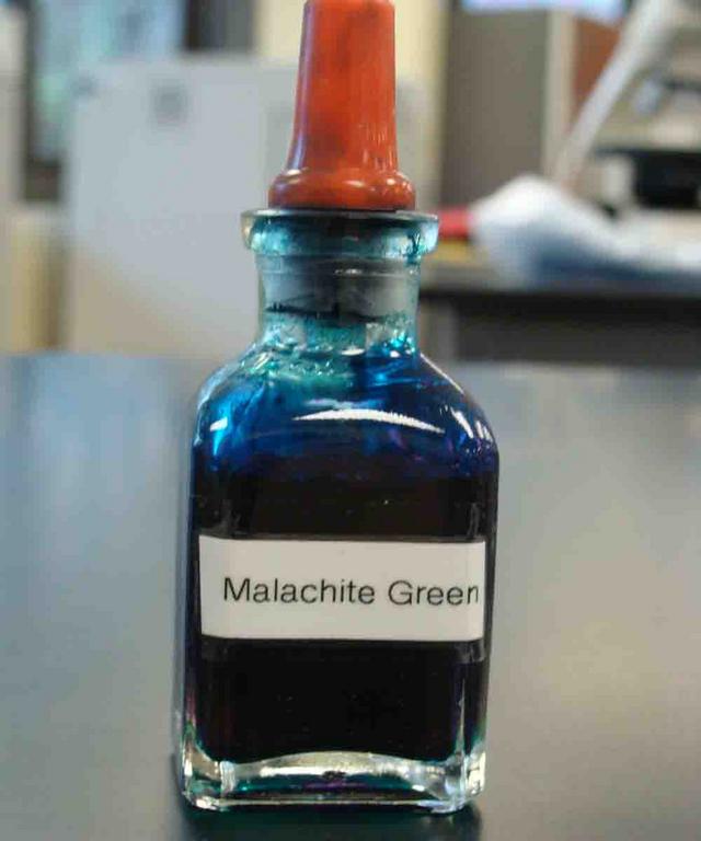
- Place the heat-fixed bacterial slide over screened water bath and then apply the primary stain malachite green.
- Allow the slide to sit over the steaming water bath for 5 minutes, reapplying stain if it begins to dry out.
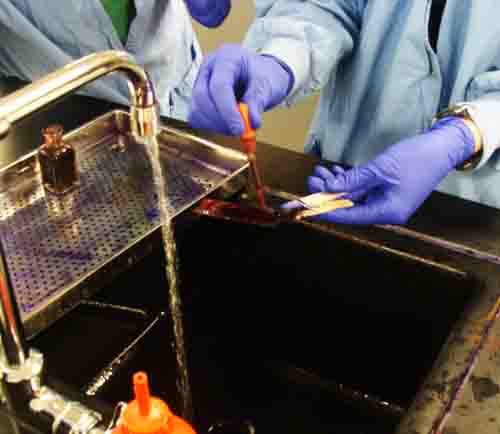
- Remove the slide from the water bath and rinse the slide with water until water runs clear.
- Flood slide with the counterstain safranin for 1 minute, then rinse.
- View specimen under oil immersion (magnification of 1000xTM) with a compound light microscope.
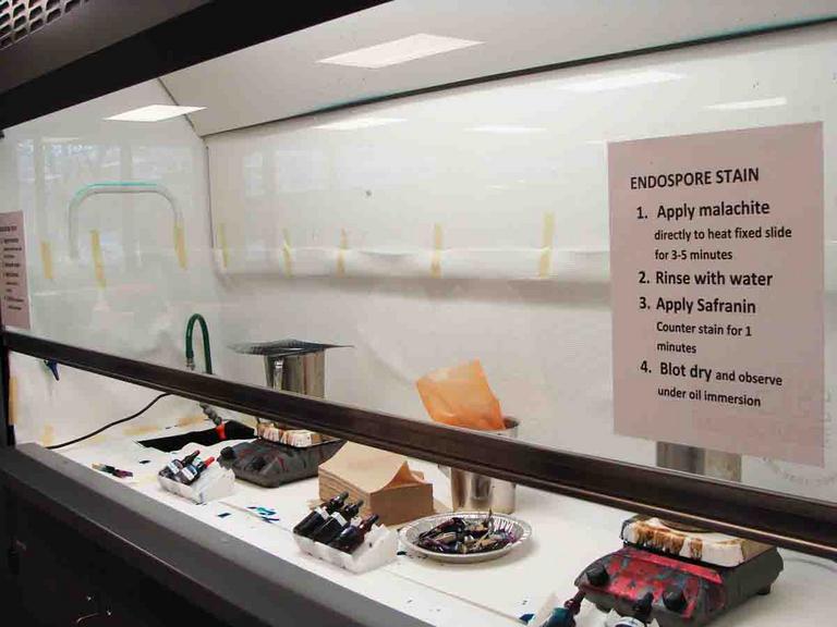
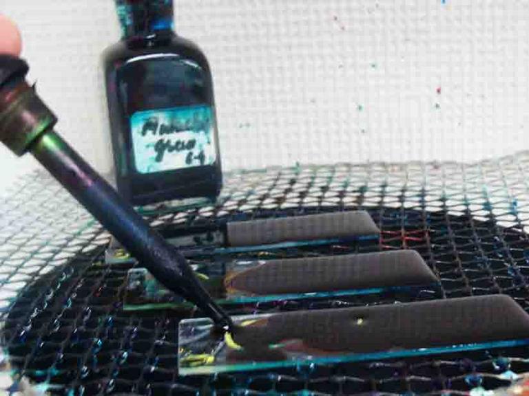
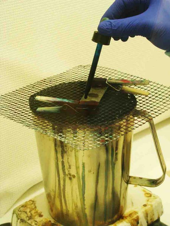
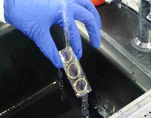
Application of Primary Stain: 1. Endospore stain set-up; 2 & 3. Malachite green being applied to slides on water bath, note in photo 3, clothes pins are used to make handling the slide easier; 4. Rinse. Make sure to rinse thoroughly enough so that there are no "chunks" of green on slide.
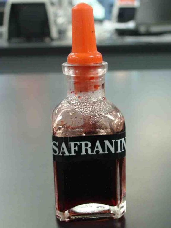
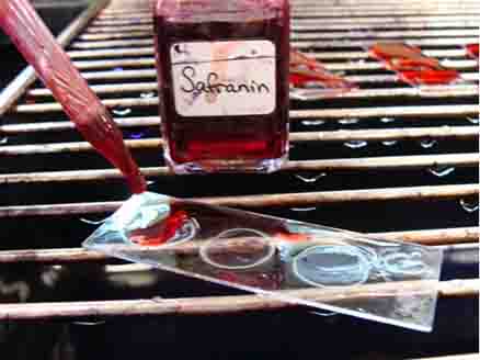

Application of Counterstain: 1. Secondary stain safranin; 2 & 3. Flood slide with safranin and leave stain on for 1 minute; 4. Rinse. Click here for more endospore stain photos.

Endospore stained bacterial smear of Bacillus subtilis under oil immersion @1000xTM. Pink rods are vegetative cells with smaller blue-green oval endospores.
SPO VIRTUAL CLASSROOMS
| ||||||
After this staining procedure, the endospores will appear green, having retained the primary stain, malachite green. Vegetative cells (bacteria are in the active, metabolizing state) will appear pink, having retained the counterstain, safranin.

Staining Bacterial Endospores
Normal water-based techniques, such as the Gram stain, will not stain these tough, resistant structures. In order to stain endspores, the dye malachite green must be forced into the spore with heat, in much the same way that carbol fuschsin is forced through the waxy mycolic acid layer of Mycobacterium in the Acid-fast stain.

SCIENCE VIDEOS

Preparing a Bacterial Sample
Prior to staining bacteria, a bacterial smear must be heat fixed onto a microscope slide. A smear is a sample of bacteria suspended in a small amount of water on a slide. That sample is then dried using heat. The heat kills the bacteria and attaches the sample to the slide so that it does not easily wash away.
When environmental conditions again become favorable, the endospore germinates, resulting in a new vegetative cell.
Bacterial smears heat fixing on the platform of a microincinerator.