 | ||||
Gram-positive Bacterial Cell Wall - P2
SPO VIRTUAL CLASSROOMS

Gram-positive Staphylococcus epidermidis @ 1000xTM+
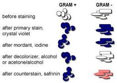
The differential Gram stain not only colors the bacteria, but the specific stain reaction distinguishes between two meaningful categories of bacteria based on the differences in their cell wall structure.
Page last updated: 11/2015
You have free access to a large collection of materials used in a college-level introductory microbiology course. The Virtual Microbiology Classroom provides a wide range of free educational resources including PowerPoint Lectures, Study Guides, Review Questions and Practice Test Questions.

PAGE 2 < Back to Page 1

End of Article
Gram Positive Bacteria
Gram+ bacteria are extremely diverse. Some are pathogens, others normal flora that is beneficial to our body. There are important groups that live in the soil and are vital to the cycling of nutrients, and yet other types of Gram+ bacteria that are used to manufacture antibiotics.
Sources & Resources
- Bauman, R. (2014) Microbiology with Diseases by Taxonomy, 4th ed.
- How to Do a Gram Stain: Identification of Gram+ and Gram- Bacteria, "Lab Notes" article from Science Prof Online.
- Differential Staining of Bacteria Laboratory Main Page.
- Gram Stain Bite Sized Tutorial: This is an extremely useful tutorial that shows, step-by-step, the Gram-staining procedure and the appearance of Gram+ and Gram- bacterial cells.
After Gram staining, Gram+ cells appear purple; their thick layers of peptidoglycan having retained the primary stain, crystal violet. Because Gram negative cells have a very thin layer of peptidoglycan layer, these cells do not retain the purple primary stain. At the end of the Gram staining procedure, Gram-negative cells retain the secondary stain, safranin, and appear pink.