 | ||||
Science Image Library - Mycobacterium Photos
Science Image Library
Mycobacterium Bacteria
from Science Prof Online

| ||||
You have free access to a large collection of materials used in a college-level introductory microbiology course. The Virtual Microbiology Classroom provides a wide range of free educational resources including PowerPoint Lectures, Study Guides, Review Questions and Practice Test Questions.

Page last updated 5/2014
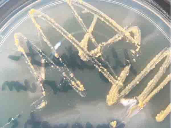
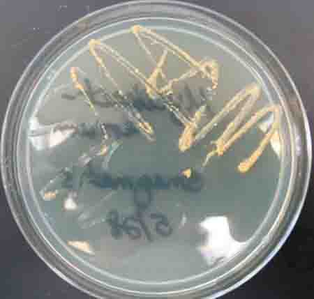
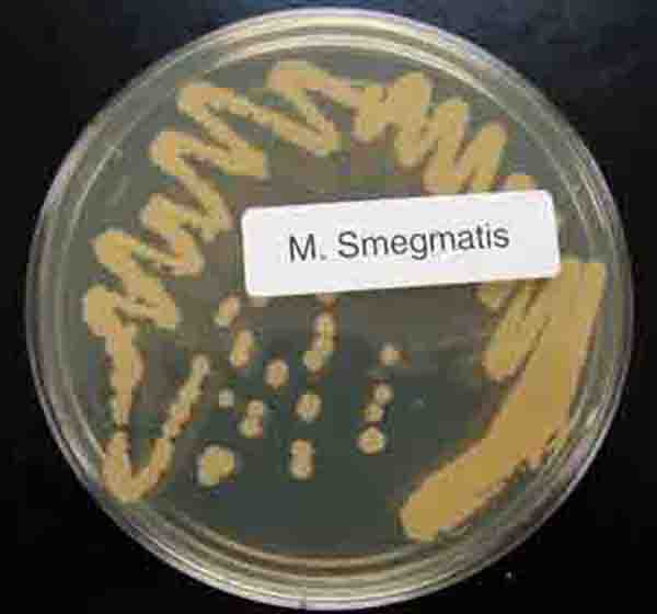
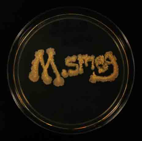
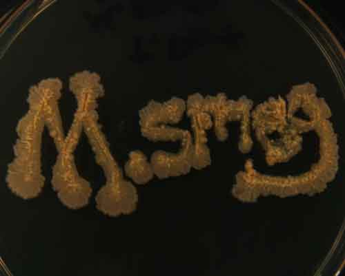
1 & 2. Streak plate of Mycobacterium smegmatis colonies growing on TSY agar, top view; 3. Streak plate of Mycobacterium smegmatis colonies growing on TSY agar, bottom view; 4 & 5. M. smeg written in the bacteria Mycobacteria smegmatis growing on TSY agar. Note, in close-up photo, the waxy appearance of the bacterial colonies.
Mycobacterium smegmatis Bacterial Colonies (Click on photo to enlarge.)
1 & 2: Acid-fast stain showing positive (Mycobacterium) results @ 1000xTM; 3 & 4. Mixed slides of acid-fast Mycobacterium (bright pink) and nonacid-fast bacteria (purple).
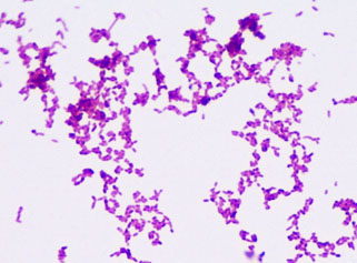
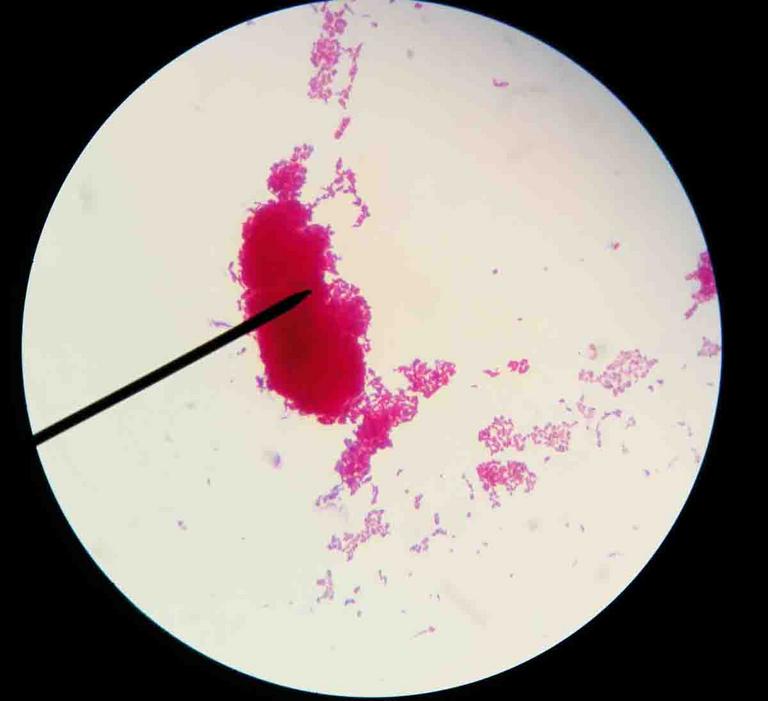
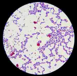
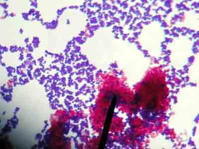
Mycobacterium Acid Fast Photos (Click on image to enlarge.)

SCIENCE PHOTOS
Didn't find what you need?
Search SPO for a Photo
SPO VIRTUAL CLASSROOMS
| ||||||
| ||||||
SPO is a FREE science education website. Donations are key in helping us provide this resource with fewer ads.
Please help!
(This donation link uses PayPal on a secure connection.)

The Microbiology Image Library is the largest photo collection on the SPO site. To help you more easily find what you're looking for, select the "See more" link of the sub-topic below that corresponds to your interests or use the search boxes.
The SPO Science Image Library is a continuously growing collection of copyright-free science photographs. If you use one of our free, low-res images, we just ask that you give us credit and provide a link to the SPO website (scienceprofonline.com). Click on photo to enlarge. To save a photo to your computer, right click on it and select "Save".
For those in need of high-resolution images, we will soon be offering hi-res files of many photos in the Science Image Library. Follow us on Twitter @ScienceProfSPO to get updates on new SPO features and products. If you need a high resolution photo now, please contact us.
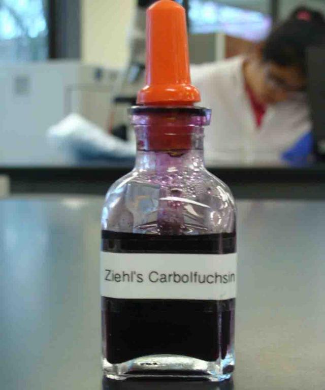
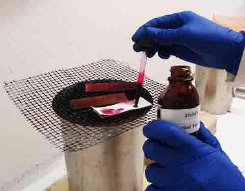

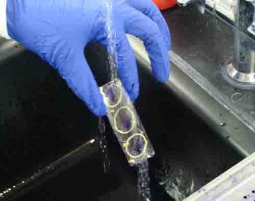


Application of Primary Stain: 1. Carbol fuchsin primary stain of acid-fat stain; 2. Carbol fuchsin being applied to slide that had been prepared with acid-fast controls and an unknown bacteria. Blotting paper has been put on top of the slide. Then the blotting paper is saturated with stain and heated over water bath; 3. Clothes pins are useful for handling the slide; 4. Blotting paper is discarded and slide is rinsed.
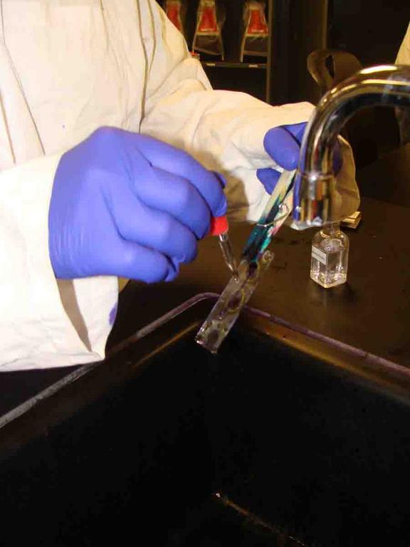
Application of Decolorizer: 1. Acid alcohol decolorizer for acid-fast stain; 2. Drizzle decolorizer down slide for 10 - 15 seconds, while watching to see that stain is removed from negative control; 3. Rinse.
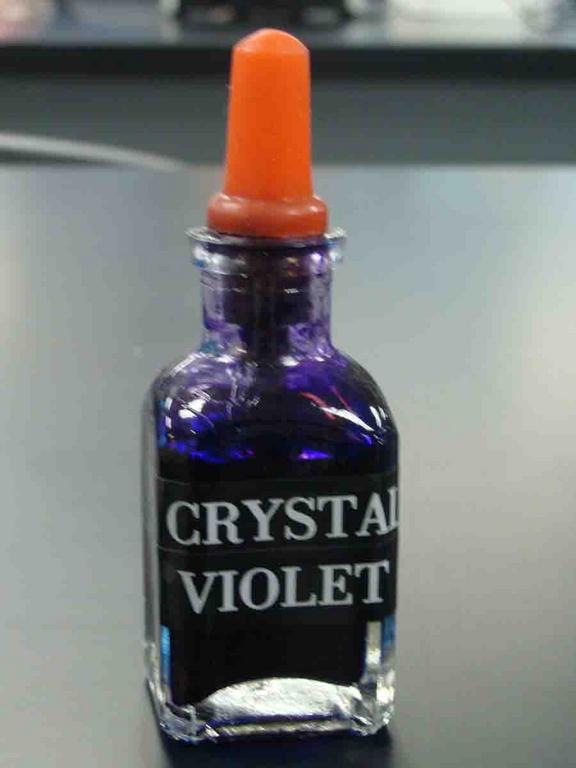
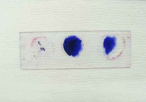
Application of Counterstain: 1. Secondary stain (coounterstain), crystal violet; 2. Crystal violet is applied to slide and left for one minute; 3. Rinse; 4. Stained acid fast slide, with + control on left, unknown in center and - control on right. Go to > Video of Acid-fast Stain.

Non-acid-fast bacteria Staphylococcus epidermidis viewed under oil immersion @ 1000xTM
Photographic Guide to Completing the Acid-fast Stain
Double click on photo strip for a slideshow of larger images.
More Acid-fast Stain Resources
- Acid-fast Ziehl Neelsen Stain Reaction: Differential Test to Identify Mycobacterium and Nocardia Bacteria, SPO Lab Notes Article.
- Identification of Unknown Bacteria Lab Exercise Main Page from the Virtual Microbiology Classroom.
- How to Use a Compound Light Microscope, SPO Lab Notes Article.
- Viewing Bacteria Using Oil Immersion Technique, SPO Lab Notes Article.