 | ||||

How to View Bacteria Under Oil Immersion
Lab Notes from Science Prof Online
Intense heat kills the microbes and firmly attaches them to the slide, so that the sample is not washed away when the sample is stained.
Article Summary: The oil immersion objective lens must be used in order to see individual bacteria through a light microscope. Here are the steps needed to get a bacterial sample in focus.
How to Use Oil Immersion to View Bacteria
You have free access to a large collection of materials used in a college-level introductory microbiology course. The Virtual Microbiology Classroom provides a wide range of free educational resources including PowerPoint Lectures, Study Guides, Review Questions and Practice Test Questions.


Oil immersion objective of a compound light microscope.

How Immersion Oil Reduces Refraction
Path of rays with immersion oil (yellow, left half) and without (right half). Rays (black) coming from specimen (red), when going through cover slip (top orange) can enter objective (dark blue) only when immersion is used. Otherwise, refraction at coverslip-air interface causes ray to miss objective.
Page last updated: 6/2015

SCIENCE PHOTOS
The easiest way to see bacteria through a microscope is to prepare a bacterial smear. A smear is a bacterial sample mixed into a drop of water on a microscope slide and then heat fixed. By placing the slide on a microincinerator tray or gently waving slide over Bunsen burner flame until dry.
SPO VIRTUAL CLASSROOMS
| ||||||

there is a total 1000X increase in the apparent size of the object being viewed. The oil immersion lens is required for viewing individual bacteria.
(Click link for downloadable pdf file)
- Place the stained bacterial smear slide in the mechanical stage apparatus. Do not use a coverslip.
- Focus the 100xTM low power objective lens on the smear and examine.
- Cover the 400xTM objective lens with a finger cot to prevent this lens from coming in contact with the oil.
- Place a drop of immersion oil directly on the sample.
- Turn nosepiece until 1000xTM objective lens snaps into position.
- Examine the lens; it should almost touch the slide and the oil should fill the space between the slide and the lens.
- While looking through the eyepiece, use only the fine adjustment knob to move the lens up and down in tiny increments; this action should bring the bacteria into focus.
- If the sample is not visible after adjustment of the fine focus, try adjusting the lighting.
- If this fails, switch back to the 10X objective lens, refocus, and try the oil immersion lens again.
Some compound microscopes also have an oil immersion objective lens, which has a magnification power of 100X. Since the magnification of the objective lenses is multiplied by the 10X magnification of the ocular lens,
| ||||

Check out our new
Sources & Resources
- Schauer Cynthia (2007) Lab Manual to Microbiology for the Health Sciences, Kalamazoo Valley Community College.
- Bauman, R. (2014) Microbiology with Diseases by Taxonomy, 4th ed., Pearson Benjamin Cummings.
- How to Use a Compound Light Microscope Lab Exercise Main Page from the Virtual Microbiology Classroom.
- How Compound Light Microscopes Magnify Objects and Are Limited by Resolution, Class Notes article from Science Prof Online.
- Differential Staining & Bacterial Controls Lab Notes Article from SPO.
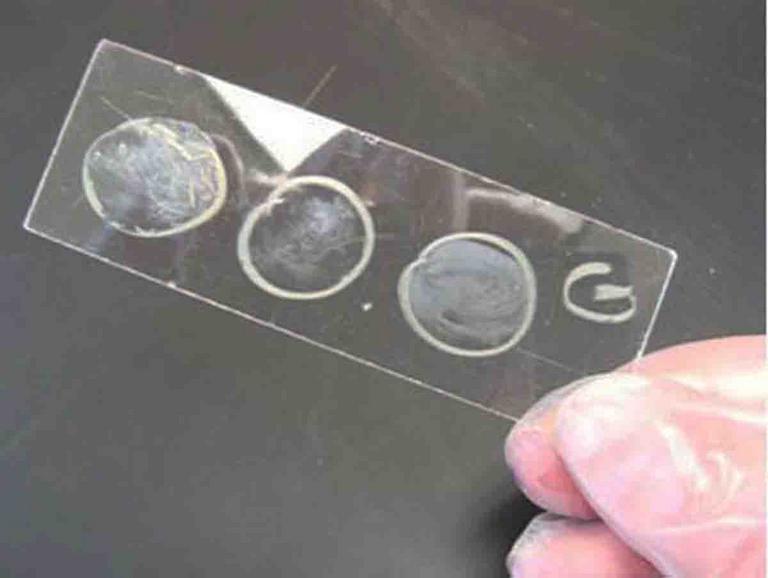
Above: Three heat fixed bacterial samples on a microscope slide.
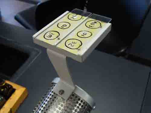
Below: Bacterial smear heat fixing on the hot platform of a miroincinerator.
The SPO website is best viewed in Microsoft Explorer, Google Chrome or Apple Safari.
The Compound Light Microscope
A microscope is considered compound when it has two sent of lenses—the ocular lenses and objective lenses. The ocular is the lens nearest the eye of the observer. The objectives are the lenses nearest the stage of the scope.
Most compound microscopes have either three or four objective lenses. If three, they most often include a:
- scanning objective, which magnifies objects 4X actual size
- low power objective, which magnifies objects 10X actual size
- high dry objective lens, which magnifies objects 40X actual size