 | ||||
How to Use a Compound Microscope
Finding and Focusing on Microscopic Objects
LAB NOTES from Science Prof Online
There are many reasons why someone using a compound scope may have difficulty seeing the specimen. Once the microscope is properly set up, and the user is sure that it’s turned on and the light source is working, there is the challenge of finding and focusing on microscopic objects. The following are solutions to problems beginners commonly encounter when using a microscope.
Article Summary: The following is a troubleshooting guide to the most common problems encountered when trying to view specimens with a compound microscope.
How to Use a Compound Light Microscope
You have free access to a large collection of materials used in a college-level introductory microbiology course. The Virtual Microbiology Classroom provides a wide range of free educational resources including PowerPoint Lectures, Study Guides, Review Questions and Practice Test Questions.
Page last updated: 10/2014
SCIENCE PHOTOS
SPO VIDEO Tutorial on Compound Light Microscope Parts & Operation
Properly Position the Specimen
Most specimens viewed with a compound light microscope are either wet-mounts of stained eukaryotic cells or bacterial smears. Make sure that the slide is properly prepared and then position it on the stage, with the area to be viewed directly over the hole in the stage where the light passes through.
 | ||||||
SPO VIRTUAL CLASSROOMS
 | ||||
Finding a Microscopic Specimen
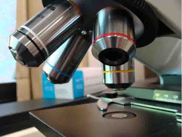
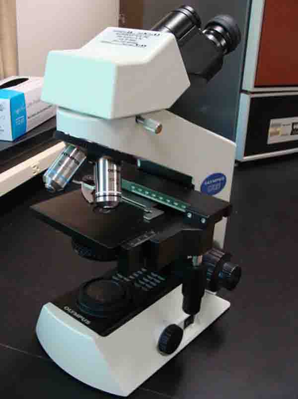
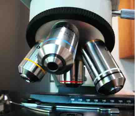
Start with the low-power objective lens: The ocular lens (nearest to the eyes) magnifies the specimen 10x. The objective lenses, attached to the nosepiece, each have a different power of magnification. The total magnification is that of the ocular lenses multiplied by the magnification of the objective lens in place. It is easiest to find objects at low magnifications, such as low power (lens with yellow band, magnified 10x), so start with one of these.
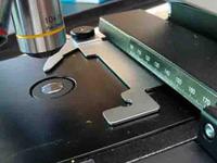
The scope may have a mechanical stage; a helpful metal apparatus used to securely position and move the slide around. The slide can be placed properly into the mechanical stage by pulling back on the claw-like clip, slipping the glass slide into the apparatus, and then letting the claw close.
Make sure the objective lens is clicked into place: Compound microscopes typically have three or four objective lenses. When the nose piece is rotated, there is an audible “click” when each lens snaps into place. If it is not clicked into position, the user will not be able to see anything.
Stage is too far from the lens: A common mistake made by those new to using a microscope is not bringing the stage close enough to the lens. When trying to focus on an object, with the scanning or low-power objective lens in place, slowly turn the coarse focus knob to bring the stage closer to the lens, until the specimen in visible. If the object being viewed is still blurry, use the fine focus to fine-tune. Never use the coarse focus adjustment with the longer objective lenses; those with a blue, or a black and white band.
Increasing Magnification
Center the specimen: As magnification increases, the field of view (area the user can see) gets smaller. If the specimen is not centered, it may no longer be visible when the user switches to a higher power lens.
Remember parfocal: Compound microscopes are parfocal, meaning that if the specimen being viewed is in focus, it will still be just about in focus when the user switches to the next higher objective lens. In other words, one doesn’t have to start from scratch focusing every time the magnification is changed. After switching to a higher power objective, manipulating the fine focus is all that should be required.
Can not find specimen after switching to higher objective power: If, after a good attempt, manipulating the fine focus does not yield a clear image of the specimen, remember the mantra “When in doubt, back it out!”; meaning switch back to the next lowest objective power, and find the specimen. Once it is in focus, try the higher power again.
More Microscope Resources
- "How big is a...?" animation from Cells Alive.
- Resolving Power Line illustration of what can be seen with different microscopes from Nobelproze.org.
- Microscopic Measurement: How Big Is that Object in the Microscope? from VirtualUrchin
- How to Use a Compound Light Microscope: Biology Lab Video Tutorial, SPO YouTube Video.
- Light Microscopy Basics animation from the Imaging Technology Group, University of Illinois.
- How Light Microscopes Magnify Objects and Are Limited by Resolution, Class Notes Article from Science Prof Online (SPO).
- Microscopic Fresh Water Pond Life Main Page from Kid Science section of Science Prof Online.
- What Is an Electron Microscope? TEM and SEM Basics, from SPO
