 | ||||
How Light Microscopes Magnify
Objects & Are Limited by Resolution
This makes the light spread out after it passes through the lens, producing an enlarged, and inverted (upside down and backwards) image of the specimen. How much the specimen is enlarged depends on the thickness of the lens, how curved the lens is, and how fast light moves through the object being viewed.
How Much Can a Light Microscope Magnify an Object?
A light microscope can clearly magnify a specimen to 1000x its actual size, when an oil immersion lens is used.
Article Summary: Light microscopes pass waves of visible radiation through lenses in order to increase the apparent size of the object being viewed. Here's are the basics.
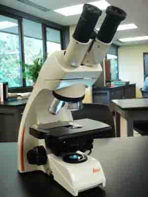
How Compound Light Microscopes Magnify Specimens
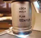
100x Objective Lens of compound light microscope provides maximum total magnification of 1000xTM.
SPO VIDEO
Tutorial on Compound Light Microscope Parts & Operation
Light waves passing through a lens bend, or refract the light. Because lenses are curved, light that passes through the edge of the lens bends more than the light that passes through the center of the lens.
SPO VIRTUAL CLASSROOMS
 | ||||||
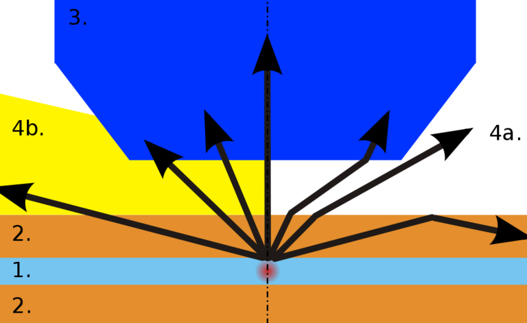
How Immersion Oil
Reduces Refraction
Path of rays with immersion oil (yellow, left half) and without (right half). Rays (black) coming from specimen (red), when going through cover slip (top orange) can enter objective (dark blue) only when immersion is used. Otherwise, refraction at coverslip-air interface causes ray to miss objective.
For some objects, such as viruses, which are typically 100nm or less in size, and the detailed ultrastructure of living cells, 1000x is not adequate magnification.
Relative Size of Microscopic Objects
Most people are, understandably, accustomed to measurements of things that are visible to the naked eye…meters, centimeters and even millimeters.
Micrometers and nanometers are units of measurement that belong solely to the microscopic world.
There are one thousand millimeters, or one hundred centimeters in a meter. The smallest object that the unaided human eye is capable of seeing is a fraction of a millimeter.
In one millimeter, there are a thousand micrometers, represented as µm (µ is the Greek lowercase letter mu). The eukaryotic cells of animals and plants are tens to hundreds of micrometers in size. Bacteria are typically just a few micrometers in size. Viruses are even smaller - mere nanometers; the next unit of smallness in the world of microbiology, with a million nanometers making up one millimeter. It is in the realm of nanometers that light microscopes lose their power to reveal clear images.
You have free access to a large collection of materials used in a college-level introductory microbiology course. The Virtual Microbiology Classroom provides a wide range of free educational resources including PowerPoint Lectures, Study Guides, Review Questions and Practice Test Questions.
Page last updated:
9/2015
The SPO website is best viewed in Google Chrome,
Microsoft Explorer or Apple Safari.
 | ||||


