 | ||||
Gram, Acid-fast & Endospore Bacterial Stains - P3
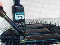
First Step of the Endospore Stain: Malachite green, the primary stain being applied slides of bacterial smears, heating over a water bath.
Endospore Staining
This stain is designed to distinguish vegetative cells of bacteria (active, living cells) from endospores. A bacterial smear is first placed over a water bath and the stain malachite green is applied. It heats on the water bath for five minutes. Then the slide is rinsed and a pink counterstain (safranin) is applied for one minute. After the final rinse, when viewing the slide under oil immersion (@1000xTM) with a compound light microscope, vegetative cells will appear pink and endospores are stained a bluish green.
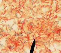
Endospore stain showing endospores (green) & vegetative cells (red) @ 1000xTM. Go to > More Endospore Stain Images
Sources and Resources
- Schauer, Cynthia (2007) Lab Manual to Microbiology for the Health Sciences, Kalamazoo Valley Community College.
- Bacterial Cell Wall & Differential Staining Lecture Main Page from the VMC
- How to Use a Compound Light Microscope, SPO Class Note Article
- Viewing Bacteria Using Oil Immersion Technique, SPO Class Notes Article
- Bauman, R. (2014) Microbiology with Diseases by Taxonomy 4th ed., Pearson Benjamin Cummings.
What the Endospore Stain Reveals about Bacteria
The endospore stain is used to identify bacteria that can produce endospores—small, dormant structures akin to “bacteria seeds.”
Forming endospores is very advantageous to the bacteria that can perform this nifty trick, such as the genera Bacilli and
Clostridium.
Endospores allow for survival under difficult environmental circumstances, including desiccation, starvation, high heat, exposure to chemicals and radiation.
Page last updated 3/2016
SPO VIRTUAL CLASSROOMS
SCIENCE VIDEOS
PAGE 3 < Back to Page 1
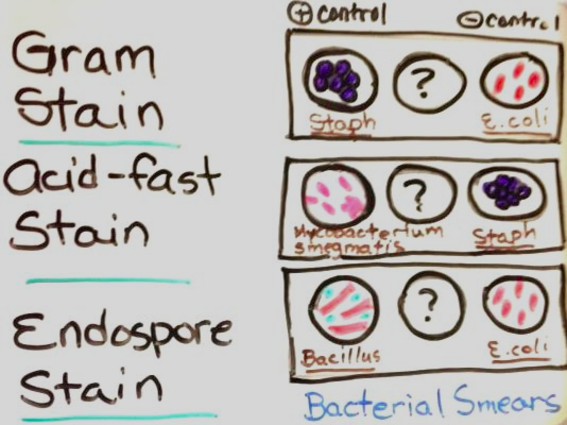
Diagram on white board of bacterial controls used for Gram, Acid-fast-and Endospore differential staining of bacterial smears. Positive control on left, negative control on right, unknown bacteria in center.
The Virtual Microbiology Classroom provides a wide range of FREE educational resources including PowerPoint Lectures, Study Guides, Review Questions and Practice Test Questions.


